

COVID-19 Screening
Through these challenging times, the Bent Neurophysiology Lab hopes to conduct research in a manner that is safe for everyone.
In addition to the necessary precautions that will be taken in the lab during face-to-face research, we ask that all participants entering the lab complete the mandatory University of Guelph COVID-19 Screening Form prior to their arrival.
Before entering the lab, please complete the following University of Guelph COVID-19 Screening Tool, and keep the email that is sent following its completion as confirmation upon your arrival.
Prior to Arrival
In addition to the above COVID-19 Screening, the Bent Neurophysiology Lab asks that you complete the COVID-19 Consent Form (attached below) prior to attending the lab.
The Bent Neurophysiology Lab team recommends that you wear comfortable clothing, preferably shorts (unless otherwise stated by your researcher). You may also choose to bring shorts, as there is washroom access in the ANNU building in which you can change upon arrival.
If you have any questions prior to arrival about the research that is being conducted, the techniques used, expectations, or COVID-19 related protocols, do not hesitate to ask. The Bent Neurophysiology Lab team is both willing and excited to perform such research in both an educational and informed manner.
Where are we Located?

There are two recommended parking lots when visiting:
- P25 which can be accessed off McGillvray St.
- P26 which can be accessed off Grange Lane.
The ANNU building has ramp and auto door access on the east side of the building which faces Gordon St., as well as elevators located on the main floor that provides access to all floors.
For walking directions from central campus to the ANNU building, click here.
The Bent Neurophysiology Lab is located at the University of Guelph in room 373 of the Animal Science and Nutrition Building (ANNU).
The address for this building is 491 Gordon Street, Guelph, ON, and is highlighted below in dark red.
What to Expect Upon Arrival
Upon arrival, parking at lots P25 and P26 behind the ANNU building is completed through a downloaded app. Upon downloading the app, each lot has a sign with an associated code that you can use to register and pay for your parking spot. Additionally, you may also choose to park in the University Center lots, which are cheaper for full-day parking.
A member of the Bent Neurophysiology Lab team will meet you at the North Loading Docks at the ANNU building, located adjacent to McGilvary St.
You can find an image of the parking lots, as well as the meeting point below.
What to Expect in the Lab
Each individual’s experience in the Lab will be very different depending on the research project that they are volunteering to be a subject for. We recommend you head over to the Ongoing Research tab, and read the study as well as what has been done so far.
Following your reading, you can return here and read about the associated laboratory technique that is being used during your visit. Hopefully, this creates an expectation regarding the procedural aspect of the lab. If you have any questions at all, do not hesitate to reach out. The Bent Neurophysiology Lab is committed to performing research in a manner that is comfortable, safe, and informative for our subjects, as well as ourselves.
Laboratory Techniques
Below are some of the laboratory techniques that are used here at the Bent Neurophysiology Lab. Associated with these techniques is a short description, and either an image or an instructional video. We hope that these descriptions build an understanding of the various techniques, as well as potentially act as an informational resource when choosing to volunteer as a subject.

Microneurography
The use of a needle electrode to record impulse trains from a single nerve fibre. The use of this needle electrode allows for direct recordings from a single afferent or efferent nerve fibre. A beneficial aspect of microneurography is that it does not require anesthetics, therefore allowing for the recording from a single nerve fibre in alert and awake human subjects.
Here at the Bent Neurophysiology Lab, we use microneurography in attempts to gain an understanding of how nerves connect in healthy individuals. We analyze this under various postural responses, functional strategies, and through interactions with other sensory input.
Galvanic Vestibular Stimulation
Galvanic Vestibular Stimulation (GVS) is a non-invasive method that uses electrical current to stimulate the eighth nerve afferents. This stimulation is completed through electrodes placed at the mastoid process of the skull, resulting in the disruption of the vestibular system.
Here at the Bent Neurophysiology Lab, we use GVS in order to create a better understanding of the relationships between sensory systems/reflexes. Specifically, we look at the role of vestibular input in “setting” the excitatory nature of the spinal cord.
In the associated video on the right, you can watch former Bent Neurophysiology Lab PhD student Adam Toth discuss GVS.

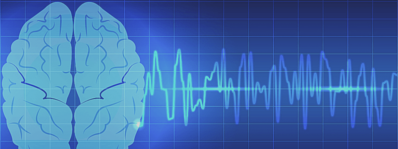
Transcranial Magnetic Stimulation
Transcranial Magnetic Stimulation (TMS) is a non-invasive method used to either excite or inhibit the descending input from the brain. This technique allows for the analyzation of the central contributions to postural reflex loops.
Here at the Bent Neurophysiology Lab, we use TMS to examine contributions from cortical areas in the control of movement.

Cutaneous Reflex Response
A cutaneous reflex response is an involuntary and often instantaneous response to a stimulus, as signalled by the cutaneous mechanoreceptors. Following the stimulus at the mechanoreceptors, the excitation results in a signal passed from afferents into the motor neuron pool of the spinal cord, where a response (involuntary, instantaneous) is initiated by an efferent signal.
Here at the Bent Neurophysiology Lab, we use cutaneous reflex responses to better understand sensory responses and neural integration.

Monofilament Testing
Monofilaments are small, thread-like objects that bend at a specific amount of force. They come in a package of many filaments that bend at different amounts of force. They are used to assist in analyzing tactile sensitivity deficits. Monofilaments specifically allow for the understanding of detection of light touch sensation.
At the Bent Neurophysiology Lab, we use monofilaments to test skin sensitivity to light touch; often before and after interventions such as heating and cooling of the skin to see if sensitivity has changed

JVP Domes
JVP Domes are small grated surfaces used for cutaneous spatial resolution management. They are used to assist in the analyzation of sensitivity deficits. JVP domes specifically allow for the understanding of spatial acuity. They contain gratings or groves, which are identified by the subjects with regards to their orientation.
In the Bent Neurophysiology Lab, we use JVP Domes to better understand spatial acuity/resolution.
In the image, the analysis of spatial acuity in the horizontal direction is shown.
Vibration
The application of vibration to the skin can be used to test the tactile sensitivity of the various mechanoreceptors present in the skin. Different frequencies of vibration are best picked up by each of the mechanoreceptors. In this way, vibration can target a certain mechanoreceptor type when testing tactile sensitivity.
In the Bent Neurophysiology Lab, we use vibration to stimulate one or more classes of mechanoreceptors in order to test the perception of the stimulus and the firing of the afferent nerves associated with these mechanoreceptors. Additionally, vibration has also recently been used in the lab in order to analyze proprioceptive feedback

Stochastic Resonance
Stochastic Resonance (SR) is a phenomenon where a tactile stimulus is enhanced by the addition of a second, subthreshold tactile stimulus (one that is too light to be detected). This enhancement can be both perceptual (it can be felt more easily) and neural (the nerve fires more strongly).
In the Bent Neurophysiology Lab, we test the capabilities of SR to enhance perception of light tactile stimuli as well as cutaneous reflex generation at the lower limb.

Electromyography
There are 2 Types of Electromyography, but both are used to record muscle activity
Surface electromyography (EMG) is a non-invasive method in which electrodes are placed on the skin to record the electrical activity of a muscle.
Indwelling EMG, similar to Surface EMG, is used to record muscle activity. However, indwelling EMG is an invasive form in which a needle electrode pierces the skin and enters directly into the muscle in order to record the electrical activity from an individual motor unit.

Electrotactile Stimulation
Electrotactile stimulation is the use of an electrical stimulus/current to evoke sensation in sensory nerves, specifically the primary afferents. Electrotactile stimulation represents an approach of achieving tactile stimulation without the physical deformation of the cutaneous mechanoreceptors.
At the Bent Neurophysiology Lab we use elecrotactile stimulation in order to evoke and analyze sensory illusions. Currently, this is specifically used to increase our understanding of the Cutaneous Rabbit Effect and the Bayesian Perceptual Model.